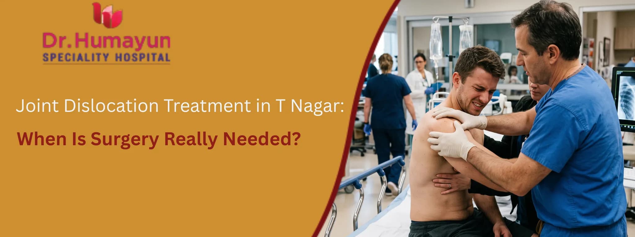

A shoulder that pops out during a fall on the cricket field. A knee that gives way and shifts under a heavy tackle. A hip that dislocates in a road traffic accident. A finger that bends the wrong way catching a thrown ball.
Joint dislocations are one of the most painful and alarming musculoskeletal injuries a person can experience. The joint looks and feels wrong. Movement is impossible. The pain is immediate and severe.
The most urgent clinical question is not whether surgery is needed. It is whether the joint can be returned to its correct position, and returned quickly, because every minute a joint remains dislocated causes progressive damage to the surrounding soft tissue, nerves, and blood vessels that the displaced bone is pressing against. This guide explains what joint dislocation treatment in T Nagar and across Chennai involves, when the non-surgical pathway is sufficient, and precisely when surgery becomes the right recommendation.
What Is a Joint Dislocation?
A dislocation is the complete separation of the bones that form a joint. Unlike a subluxation, where the bones are partly displaced but maintain some contact, a full dislocation means the joint surfaces have lost all contact with one another.
The key characteristics of a dislocated joint include:
-
Severe, immediate pain at the joint
-
Visible deformity or the joint looking noticeably out of place
-
Complete inability to move the joint normally
-
Swelling and bruising developing rapidly
-
In some cases, numbness or tingling if a nerve is being compressed by the displaced bone
A dislocation can affect any joint in the body. The most commonly dislocated joints in clinical practice are:
-
Shoulder (glenohumeral joint) : the most frequently dislocated joint overall
-
Finger (interphalangeal joints) : particularly common in ball sports
-
Elbow
-
Patella (kneecap)
-
Hip
-
Knee (tibiofemoral): rare but serious, associated with vascular injury risk
Each joint has a specific anatomy, specific mechanisms of dislocation, specific associated injury risks, and specific criteria for deciding between conservative and surgical management.
The Emergency First Step, Reduction
The most urgent priority in joint dislocation treatment in Chennai is reduction: returning the dislocated joint to its correct position. This is performed by an orthopaedic specialist, not attempted at home.
Attempting to self-reduce a dislocation, or having an untrained person attempt it, risks fracturing the displaced bone, damaging the surrounding blood vessels, or damaging the cartilage of the joint surface. It also causes severe additional pain and can make formal reduction significantly more difficult.
In the emergency or orthopaedic setting, reduction is performed with:
-
Local anaesthetic, conscious sedation, or general anaesthesia depending on the joint and the degree of muscle spasm
-
Specific traction and rotation manoeuvres that guide the displaced bone back into its socket
-
X-ray imaging before reduction to confirm the dislocation pattern and identify any associated fracture
-
X-ray after reduction to confirm the joint is correctly positioned and no fracture has occurred during the procedure
In a clinical audit of 100 patients presenting with shoulder dislocations, closed reduction under anaesthesia was successful in 98% of cases. Open surgical reduction was required in only 2%, specifically in cases where soft tissue, including tendons or nerves, had become interposed in the joint space and prevented closed reduction.
When Non-Surgical Treatment Is Sufficient
After successful closed reduction, most joint dislocations are managed conservatively without surgery. Non-surgical joint dislocation treatment in T Nagar involves immobilisation, pain management, and structured rehabilitation.
Immobilisation
After reduction, the joint is immobilised in a position that protects the capsule and ligaments while they begin to heal:
-
Shoulder: Sling for two to three weeks for most first-time dislocations, with early movement exercises introduced gradually under physiotherapy supervision
-
Finger: Buddy taping or a splint for two to three weeks, with early range of motion encouraged to prevent stiffness
-
Patella: Knee brace with the leg extended for two to four weeks, followed by progressive flexion
-
Elbow: Above-elbow backslab for one to two weeks, with early movement initiated to prevent the stiffness that elbows develop rapidly with prolonged immobilisation
-
Hip: Activity restriction and avoidance of positions that risk redislocation for six to twelve weeks
Rehabilitation
Rehabilitation is not optional after a joint dislocation. It is the component that determines long-term joint stability and function. Regardless of whether the patient was also offered surgery, physiotherapy that rebuilds the muscle strength around the reduced joint is what prevents redislocation and restores full function.
After a shoulder dislocation specifically, the goal of rehabilitation is to strengthen the rotator cuff muscles that hold the humeral head (ball of the shoulder) against the glenoid (socket). Without this muscle rehabilitation, even a joint that was correctly reduced and properly immobilised will remain vulnerable to redislocation.


When Surgery Is Recommended for Joint Dislocation
This is the central clinical question the blog is designed to answer. Surgery is not needed for every dislocation, but there are specific, well-defined clinical situations where conservative management is insufficient and surgical intervention changes the long-term outcome.
When Closed Reduction Cannot Be Achieved
A small proportion of dislocations cannot be reduced by closed manipulation. This occurs when a structure, typically a tendon or torn capsule, becomes interposed between the joint surfaces and physically prevents the bone from returning to its socket. Open surgical reduction is then required to clear the obstruction and reduce the joint under direct visualisation.
When There Is an Associated Fracture
A dislocation associated with a fracture of the joint surfaces or surrounding bone, called a fracture-dislocation, frequently requires surgical management. The fracture fragment may prevent stable joint reduction, compromise the joint surface in a way that requires anatomical realignment, or carry a blood supply risk that needs surgical attention.
Common examples include:
-
Shoulder dislocation with a greater tuberosity fracture or significant Hill-Sachs defect
-
Hip dislocation with an acetabular or femoral head fracture
-
Elbow dislocation with a radial head fracture (terrible triad injury)
-
Knee dislocation with multiligamentous injury or bony avulsion
When There Is Significant Soft Tissue Damage
A dislocation that has torn a major ligament, capsule, or tendon may produce a joint that remains unstable even after successful reduction. Surgery to repair the torn structure is then required to restore joint stability.
In the shoulder, this includes:
-
A Bankart lesion, where the anterior labrum (cartilage rim of the socket) is torn from the glenoid, producing the classic instability seen after anterior shoulder dislocations. Arthroscopic Bankart repair restores the labrum to the glenoid rim and significantly reduces redislocation risk.
-
A large Hill-Sachs lesion, where a dent is created in the humeral head by impaction against the glenoid at the time of dislocation, may require a bone grafting procedure (Latarjet procedure) when the bone loss compromises joint stability.
Recurrent Dislocation and Instability
This is the most common surgical indication in joint dislocation treatment in Chennai, particularly for the shoulder.
Young patients under 35 who experience a first-time traumatic shoulder dislocation face a redislocation rate of 70 to 90% without surgical stabilisation. Older patients have a lower redislocation rate, which is why the surgical recommendation for a first-time dislocation varies by age.
For patients who have experienced multiple dislocations and continue to experience redislocation or ongoing joint instability despite rehabilitation, surgery is the definitive treatment. Recurrent instability may require surgical treatment. Conservative management of recurrent instability produces progressively worsening results with each redislocation episode as the labrum, capsule, and bone are further damaged.
Specific High-Risk Dislocations
Certain dislocation types carry specific associated injury risks that may require urgent surgical assessment:
-
Knee (tibiofemoral) dislocation: This is a high-velocity, high-energy injury associated with potential rupture of the popliteal artery in up to 20 to 30% of cases. Urgent vascular assessment is mandatory after any knee dislocation. Surgical vascular repair may be needed alongside multiligamentous reconstruction.
-
Hip dislocation: The blood supply to the femoral head is at risk following hip dislocation. Avascular necrosis (bone death from loss of blood supply) is a recognised complication, and the risk increases the longer the hip remains unreduced. Prompt reduction is the most important prognostic factor. Associated acetabular fractures require surgical management.
-
Open dislocations: When the skin is broken over the dislocation site, this constitutes a surgical emergency requiring immediate debridement and repair.
The Consequences of Delayed or Inadequate Treatment
A dislocation that is not reduced promptly, or that is inadequately treated after reduction, carries specific long-term consequences:
-
Avascular necrosis: Particularly in the hip, where the blood supply to the femoral head traverses vessels that are vulnerable to disruption when the hip is dislocated
-
Post-traumatic arthritis: Cartilage damage at the time of dislocation accelerates joint degeneration, particularly if the joint surface was impacted or if reduction was delayed
-
Chronic instability: An unreduced or repeatedly dislocating joint develops progressive capsular and labral damage that makes each subsequent dislocation easier and surgical correction more complex
-
Nerve damage: Prolonged pressure from the displaced bone on surrounding nerves produces neuropraxia that may not fully recover if treatment is delayed
-
Stiffness: Immobilisation without appropriate early mobilisation and physiotherapy, particularly in elbow and finger dislocations, leads to significant joint stiffness that can be permanent
Meet Dr. Omer Sheriff: Best Orthopaedist in T Nagar for Dislocation Management
For joint dislocation treatment in T Nagar and across Chennai, Dr. Omer Sheriff at Humayun Hospital provides the full clinical pathway from acute reduction to surgical stabilisation where needed.
Dr. Omer Sheriff is a Consultant Orthopaedic and Joint Replacement Surgeon at Humayun Hospital, T Nagar, Chennai. He holds an MBBS, Diploma in Orthopaedics (Sri Ramachandra University), MS in Orthopaedics, and a Fellowship in Joint Replacement. He trained at CMC Vellore and brings international experience from Hamad Medical Corporation, Doha, Qatar, to his clinical practice.
With over 25 years of experience and more than 3,500 successful orthopaedic procedures, his expertise in joint dislocation treatment in Chennai covers:
-
Closed reduction under anaesthesia for acute shoulder, elbow, patella, hip, and finger dislocations
-
Imaging assessment including X-ray, CT, and MRI for associated fractures and soft tissue injuries
-
Arthroscopic Bankart repair and Latarjet procedure for shoulder instability
-
Multiligamentous knee reconstruction after complex knee dislocations
-
Surgical fixation for fracture-dislocations across all joint types
-
Physiotherapy coordination and rehabilitation planning post-reduction and post-surgery
Dr. Omer Sheriff is a life member of the Indian Orthopaedic Association (IOA), the Indian Society of Hip and Knee Surgeons (ISHKS), and the Tamil Nadu Orthopaedic Association (TNOA).
Chat with our medica care assistant for quick guidance and support.