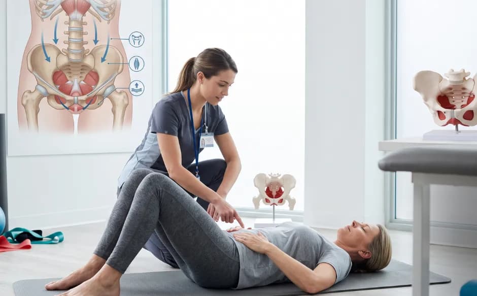

That sudden, sharp pain shooting from your lower back deep into your leg is not something you imagined, and it is certainly not something you simply have to endure. Commonly known as a slip disc, a lumbar disc herniation occurs when the soft, gel-like shock absorber between your vertebrae bulges or tears, placing direct, irritating pressure on nearby spinal nerve roots.
The lower back carries the full mechanical weight of the upper body, making the lowest spinal segments exceptionally vulnerable to structural wear, progressive degeneration, and sudden injury. Fortunately, a slip disc is not an automatic ticket to the operating room.
With over 90% of cases resolving successfully through structured, non-surgical therapies, the key to a complete recovery lies in accurate tracking and timely intervention. This guide breaks down the defining symptoms of a lumbar slip disc, details essential diagnostic imaging, and explores evidence-backed treatment options designed to restore your pain-free mobility.
What Is a Slip Disc and Why Does It Happen in the Lower Back?
A slip disc, more precisely known as a disc herniation or disc prolapse, occurs when the soft inner material of an intervertebral disc pushes through a tear in its tougher outer casing. When this protruding material presses against a nearby spinal nerve root, it produces the pain, numbness, and weakness that make slip disc one of the most debilitating musculoskeletal conditions in everyday clinical practice.
The lower back, specifically the lumbar spine, is where slip discs occur most frequently. As AAOS OrthoInfo confirms, disc herniation most often happens in the lower back (lumbar spine) and is a common cause of both lower back pain and leg pain. An estimated 60 to 80% of people experience significant lower back pain at some point in their lives, and a herniated disc is among the most common structural causes.
The reason the lower back is most vulnerable is biomechanical. The lumbar spine carries the full weight of the upper body and is the pivot point for almost every movement we make, from sitting and bending to lifting and rotating. The L4-L5 and L5-S1 disc levels, the two lowest discs in the lumbar spine, absorb the greatest mechanical load and are therefore the most frequent sites of herniation.
Structural Progression of a Disc Herniation
Each intervertebral disc has two components. The outer ring, called the annulus fibrosus, is a tough, layered structure of fibrocartilage. The inner core, called the nucleus pulposus, is a soft, gel-like material that acts as a shock absorber, distributing pressure evenly across the vertebrae above and below it.
Disc herniation occurs in stages. First, repeated stress or acute injury causes small tears in the annular fibres. As the annulus weakens, the nucleus pulposus begins to push outward. In a disc bulge, the annulus remains intact but balloons outward. In a true herniation, the nucleus breaks through the outer ring entirely. In the most severe cases, sequestration, a fragment of nucleus material separates completely and floats freely in the spinal canal.
Each stage places increasing pressure on the adjacent nerve roots. The specific nerve root being compressed determines exactly which symptoms a patient experiences and in which part of the leg or foot those symptoms appear.
Slip Disc Symptoms in the Lower Back: What to Look For
Slip disc symptoms in the lower back vary depending on the disc level involved and the degree of nerve compression. Understanding the symptom pattern helps both the patient and the doctor identify the likely location of the herniation before imaging confirms it.
-
Lower back pain is often the first symptom, frequently described as a deep ache or sudden severe pain that worsens with bending forward, sitting for extended periods, coughing, or sneezing. These activities all increase the internal pressure within the disc, forcing more nuclear material against the nerve.
-
Leg pain (sciatica) is the defining symptom of lumbar disc herniation involving nerve root compression. As Penn Medicine describes, symptoms include sharp pain in one part of the leg, hip, or buttocks and numbness in other parts, as well as pain or numbness on the back of the calf or sole of the foot.
-
Numbness and tingling in the leg, foot, or specific toes are caused by compression disrupting the sensory signals travelling through the affected nerve root.
-
Muscle weakness in the leg or foot may develop when motor nerve fibres are compressed. Weakness in lifting the foot (foot drop), difficulty standing on tiptoe, or reduced knee reflex are each specific indicators of which disc level is involved.
-
Pain that is worse at night or with prolonged sitting is characteristic of slip disc, as lying in certain positions or sitting increases nerve root tension.
Importantly, symptoms almost always affect one side of the body. Bilateral leg symptoms, or symptoms involving bowel and bladder function, are a red flag requiring immediate assessment.
The Sciatica Connection: When Pain Travels Down the Leg
The term sciatica describes radiating leg pain caused by irritation or compression of the sciatic nerve, which is formed by the L4, L5, S1, S2, and S3 nerve roots. Because lumbar disc herniations most commonly involve L4-L5 or L5-S1, sciatica is the most frequent presenting symptom of a lower back slip disc.
L4-L5 herniation compresses the L5 nerve root, typically causing pain and numbness running from the outer hip down the outer shin to the top of the foot and big toe, and weakness in lifting the foot upward (dorsiflexion). A specific 2025-confirmed clinical finding for L5 radiculopathy is a Trendelenburg gait, where the hip drops on the affected side during single-leg stance, indicating gluteus medius weakness.
L5-S1 herniation compresses the S1 nerve root, causing pain and numbness along the back of the thigh, calf, and outer foot, with weakness in standing on tiptoe and a reduced or absent ankle reflex.
As NCBI InformedHealth explains, a slipped disc in the lumbar region causes the spinal disc to press against nerves, resulting in severe pain that can radiate down the leg. The location of that leg pain is a precise anatomical map of which nerve is being compressed.
Emergency Symptoms That Cannot Wait
While most slip disc presentations are painful but not urgent, one symptom cluster demands same-day emergency evaluation. Do not wait for a scheduled appointment if you develop:
-
Loss of bladder or bowel control, or difficulty urinating
-
Numbness in the groin, inner thighs, or around the anus (saddle anaesthesia)
-
Sudden, severe weakness in both legs
-
Progressive neurological deficit affecting multiple muscle groups
These symptoms indicate cauda equina syndrome, a condition where a large herniation compresses multiple nerve roots simultaneously. Without emergency surgical decompression, permanent loss of bowel, bladder, and lower limb function can result. This is a spinal surgical emergency.
What Causes a Slip Disc in the Lower Back?
Slip discs rarely result from a single dramatic event. They develop over time and are then triggered by an incident that crosses the disc's tolerance threshold. The most common contributing factors include:
-
Age-related disc degeneration: Disc water content decreases with age, making the nucleus less gel-like and the annulus more brittle. Degenerated discs are significantly more prone to herniation under normal loading. This process begins in the late 20s and accelerates with each decade.
-
Heavy or improper lifting: Lifting heavy objects while bending the spine (rather than using the legs) dramatically increases compressive and shear forces on lumbar discs, particularly at L4-L5 and L5-S1.
-
Prolonged sitting and sedentary lifestyle: Sitting places approximately 40% more compressive force on lumbar discs than standing. Urban desk-based work environments in Indian cities create sustained disc loading across the working day.
-
Repetitive bending and twisting: Occupations and activities involving repeated forward bending, twisting, or vibration (such as driving for extended periods) progressively fatigue the annular fibres.
-
Obesity: Excess body weight increases the baseline compressive load on lumbar discs, accelerating both degeneration and the risk of herniation.
-
Genetic factors: Research confirms that a family history of disc disease significantly increases an individual's risk, independent of occupational or lifestyle factors.


How a Slip Disc in the Lower Back Is Diagnosed Accurately
Diagnosis combines clinical assessment with targeted imaging, and both components are necessary for accurate management planning.
Clinical examination is the starting point. The doctor assesses lower back range of motion, performs a straight leg raise test (the most sensitive clinical test for lumbar nerve root compression), evaluates muscle power, sensation, and reflexes in both legs, and identifies the neurological level of any deficit.
MRI (Magnetic Resonance Imaging) is the gold standard investigation for lumbar disc herniation. As Cleveland Clinic confirms, MRI provides detailed images of spinal discs and nerve compression, making it the most informative single investigation for slip disc assessment. One important caveat: many people have disc herniation visible on MRI without experiencing any symptoms. Diagnosing the source of pain requires matching MRI findings with clinical symptoms, not relying on imaging alone.
X-ray is performed to exclude fractures, assess spinal alignment, and identify bony abnormalities contributing to symptoms.
CT scan provides cross-sectional views of the spine and is useful when MRI is contraindicated or when bony detail is specifically needed.
Electromyography (EMG) and nerve conduction studies measure nerve function and identify the degree of nerve damage, particularly when symptoms are of longer duration or neurological deficits are significant.
Treatment Stage 1: Conservative Management
The critical starting point for nearly every patient is this: as Cleveland Clinic confirms, most slip disc cases resolve on their own within four to six weeks. Over 90% of patients recover fully without surgical intervention when treated appropriately.
Conservative management includes:
-
Activity modification: Avoiding the positions and activities that worsen symptoms while maintaining as much normal movement as possible. Prolonged bed rest is not recommended. Staying active, within pain tolerance, is associated with faster recovery.
-
Pain medication: NSAIDs (ibuprofen, diclofenac) reduce disc-related inflammation and nerve root irritation. Muscle relaxants manage associated paraspinal muscle spasm. Neuropathic pain medications such as pregabalin and gabapentin specifically address the shooting, burning nerve pain of sciatica.
-
Physiotherapy and targeted exercise: A structured physiotherapy programme addresses core muscle strengthening, lumbar stabilisation, and nerve mobilisation techniques. As Patient,info notes, staying active is crucial for recovery, with people who remain active recovering more quickly and being less likely to develop persistent back pain.
-
Hot and cold therapy: Ice packs in the acute phase reduce inflammation. Heat application in the subacute phase eases muscle spasm and improves tissue flexibility.
Treatment Stage 2: Interventional Pain Management
When conservative management does not achieve adequate pain relief within 6 to 8 weeks, interventional procedures provide targeted treatment with a significantly higher response rate than oral medications alone.
-
Epidural steroid injections (ESI): A precisely guided injection of corticosteroid into the epidural space adjacent to the compressed nerve root delivers anti-inflammatory medication directly to the site of nerve irritation. As UPMC confirms, epidural steroid injections reduce inflammation and relieve pain for weeks to months, allowing patients to engage more effectively with physiotherapy.
-
Nerve root blocks: A targeted injection around a specific nerve root both confirms the diagnosis (if the injection reproduces and then relieves the patient's pain) and provides therapeutic relief.
-
Intradiscal procedures: For specific cases of painful disc degeneration without significant nerve compression, procedures such as radiofrequency ablation of disc pain generators may be considered.
Treatment Stage 3: Surgery, When It Becomes Necessary
Surgery is indicated for a minority of patients: those with persistent, disabling symptoms after 6 to 12 weeks of appropriate conservative and interventional management, those with progressive neurological deficits (worsening foot drop, increasing weakness), and those presenting with cauda equina syndrome as an emergency.
Microdiscectomy is the most commonly performed procedure for lumbar disc herniation. Through a small incision, a portion of the herniated disc material pressing on the nerve root is carefully removed under magnification. As AAOS OrthoInfo confirms, this surgery reliably relieves leg pain caused by nerve compression, with most patients experiencing significant improvement.
Endoscopic discectomy is a minimally invasive alternative using a narrow endoscope inserted through a very small incision. It offers comparable clinical outcomes with reduced tissue disruption, shorter hospital stay, and faster return to normal activity.
Spinal fusion is reserved for cases where disc herniation is associated with significant spinal instability or where multiple level disease requires structural support after decompression.
Importantly, research published in Frontiers in Medicine (July 2025) documented the phenomenon of spontaneous reabsorption of herniated disc material, particularly in large herniations, where the immune system gradually reabsorbs the protruding disc material over months. This is why the decision to proceed to surgery requires patience, systematic conservative management, and careful clinical monitoring rather than an early surgical decision.
Recovery and Preventing Recurrence
Most patients who undergo microdiscectomy or endoscopic discectomy return to light activity within 1 to 2 weeks and resume normal daily life within 4 to 6 weeks. Full return to strenuous activity or heavy physical work typically takes 8 to 12 weeks.
Long-term prevention of recurrence focuses on:
-
Core strengthening and lumbar stabilisation exercises maintained as a permanent habit
-
Correct lifting technique at all times (bend the knees, keep the spine neutral, hold the load close to the body)
-
Ergonomic adjustments to the workstation including a supportive chair, monitor at eye level, and regular standing breaks
-
Healthy body weight to reduce baseline disc loading
-
Avoiding prolonged sitting without breaks, particularly during long commutes
Orthopaedic and Spine Care at Dr. Humayun Speciality Hospital, T. Nagar, Chennai
At Dr. Humayun Speciality Hospital, the orthopaedic and spine team provides comprehensive assessment and management of lumbar disc herniation across all stages of the condition, from the first presentation with acute back and leg pain through to post-operative rehabilitation.
Services include clinical neurological assessment of radiculopathy, in-house MRI and imaging for precise disc level identification, physiotherapy and pain management for conservative treatment, specialist orthopaedic consultation for interventional and surgical decision-making, and post-operative rehabilitation programmes.
The team's approach is structured around one priority: matching the right treatment to the right stage of the condition, with surgery reserved for those who genuinely require it.
Stop Managing It. Start Treating It.
Lower back and leg pain from a slip disc does not have to define your daily life. With the right diagnosis and the right treatment plan at the right stage, the vast majority of people return to full, pain-free activity without surgery.
Dealing with lower back pain, sciatica, or numbness in the leg that has not improved?
Chat with our care assistant for quick guidance and support and book an orthopaedic assessment at Dr. Humayun Speciality Hospital, T. Nagar, Chennai.