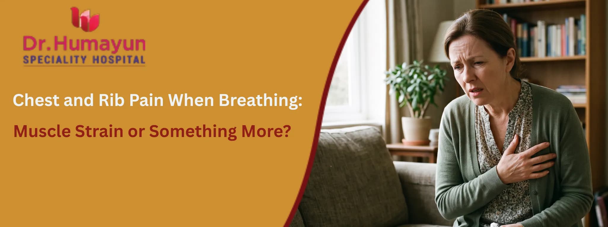

A sharp jab when you take a deep breath. An ache along the ribs that worsens every time you cough, sneeze, or reach sideways. A tightness across the chest that makes taking a full breath genuinely uncomfortable.
For most people who experience chest and rib pain when breathing, the instinct is to assume a pulled muscle. A hard gym session. A bad cough last week. An awkward movement lifting something heavy. And in many cases, that assumption is correct.
But chest pain when breathing is one of the symptoms in medicine where the overlap between benign and serious causes is most dangerous. A pulled intercostal muscle and a pulmonary embolism can both produce sharp, breath-dependent pain. The causes and outcomes are dramatically different.
Understanding how to distinguish muscular chest pain from pain that warrants urgent evaluation is the practical knowledge this guide is designed to provide.
How Breathing Produces Chest and Rib Pain
To understand why so many different conditions produce chest pain with breathing, it helps to understand what happens mechanically when we breathe.
With each breath in, the intercostal muscles (between the ribs), the diaphragm, and the chest wall muscles expand the ribcage. The lungs expand inside the chest cavity. The pleura, a thin two-layered membrane lining both the lungs and the inside of the chest wall, slides smoothly against itself. Every structure in and around the chest moves with each breath.
This means any inflamed, damaged, or irritated structure in the chest, whether muscle, cartilage, bone, membrane, or lung tissue, is subjected to movement and pressure with every single breath. Pain that would be manageable at rest becomes sharp and constant because breathing cannot be stopped.
Common Causes of Chest and Rib Pain When Breathing
Intercostal Muscle Strain
The intercostal muscles run between each pair of adjacent ribs and are the primary muscles of respiration. When they are strained, overstretched, or torn, the resulting pain is felt along the ribs and is directly worsened by breathing, coughing, sneezing, twisting, and reaching.
Intercostal strain can happen during:
-
Heavy lifting or gym exercise, particularly rowing, bench press, or any overhead movement
-
Sports involving twisting or throwing
-
A sudden, forceful cough or several days of severe coughing
-
A direct blow to the chest wall
-
An awkward movement during sleep or getting in or out of a vehicle
The defining clinical feature of intercostal muscle strain is that pressing directly on the painful area reproduces the exact same pain. The tenderness is localised, often to one specific interspace between ribs, and the pain is described as sharp or burning rather than pressure-like or crushing.
Mild intercostal strains heal within one to three weeks with rest, modified activity, and anti-inflammatory pain relief. More significant strains may take four to six weeks.
Costochondritis
Costochondritis is inflammation of the cartilage that connects the ribs to the breastbone (sternum). It produces a sharp or aching pain that is felt at the front of the chest, specifically where the ribs join the sternum, and that worsens with deep breathing, coughing, or pressure on the area.
Costochondritis worsens when taking a deep breath, coughing, or sneezing, or with any movement of the chest wall, and it often affects more than one rib. It is more common on the left side of the breastbone, which is one of the reasons it is frequently mistaken by patients for heart pain.
The key distinguishing feature is reproducibility: pressing on the costochondral junctions at the front of the chest triggers the same pain the patient reports with breathing. This is the single most useful clinical sign in differentiating costochondritis from cardiac pain.
Costochondritis often resolves without specific treatment over several weeks. NSAIDs (ibuprofen) reduce inflammation and pain during recovery. In persistent or severe cases, a short course of oral steroids or a local corticosteroid injection may be used.
Rib Fracture
A fractured rib produces severe, sharply localised chest and rib pain when breathing. It follows a specific mechanism, typically direct chest wall trauma such as a fall, a road accident, a sports collision, or in older adults with osteoporosis, even a forceful cough.
Features suggesting a rib fracture rather than simple muscle strain:
-
A clear traumatic event preceding the pain
-
Pain that is extremely severe with even minimal breathing
-
A visible or palpable deformity of the rib
-
In multiple rib fractures, paradoxical movement of the chest wall (a section that moves inward rather than outward during inspiration), which is a medical emergency
Simple rib fractures without displacement heal over four to six weeks. Pain management, including adequate analgesia to allow normal breathing, is critical because inadequate pain control causes shallow breathing and increases the risk of pneumonia.
Pleurisy
Pleurisy is inflammation of the pleural membranes lining the lungs and chest cavity. When inflamed, the two layers rub against each other during breathing rather than sliding smoothly, producing a sharp, stabbing pain that is directly worse with inspiration and often with coughing.
Pleurisy is a sign of another underlying process rather than a primary diagnosis. It is caused by viral or bacterial infections, pulmonary embolism, autoimmune conditions including lupus, pneumonia, and inflammatory lung conditions.
The pain of pleurisy is often described as knife-like and worsens dramatically with a full breath. Patients typically take shallow breaths instinctively to avoid pain. Unlike muscular pain, pleurisy is not reproduced by pressing on the chest wall and does not vary with posture or movement in the same way intercostal pain does.
The American Academy of Family Physicians emphasises that pulmonary embolism is the most common life-threatening cause of pleuritic chest pain and should be considered in all patients with pleuritic symptoms. Pleurisy always warrants clinical assessment.
Pneumonia
Pneumonia causes inflammation and consolidation of lung tissue, producing chest and rib pain when breathing alongside fever, productive cough, breathlessness, and general illness. The pain is typically felt over the affected lobe and worsens with each breath as the inflamed lung moves.
The pain of pneumonia is accompanied by systemic illness, fever, and respiratory symptoms that distinguish it from purely muscular pain. Pneumonia requires antibiotic treatment and in moderate to severe cases, hospitalisation.


Pulmonary Embolism
A pulmonary embolism (PE) is a blood clot in the pulmonary arteries that produces sudden-onset chest and rib pain when breathing, often described as pleuritic, alongside sudden breathlessness, rapid heart rate, and in some cases coughing up blood or dizziness.
PE is life-threatening. It is the most important condition to exclude in any patient presenting with sudden breathing-related chest pain and breathlessness, even when the pain seems localised to one area. A patient who has recently been immobile, who has had recent surgery or a long flight, or who has a known clotting disorder is at elevated risk.
Pericarditis
Pericarditis is inflammation of the sac surrounding the heart. It produces chest pain that is typically sharp, worsened by breathing and by lying down, and improved by sitting forward. It may be associated with a low-grade fever and a recent viral illness.
The positional nature is a key distinguishing feature: pericarditis pain is often significantly better when the patient leans forward. This is unusual for muscular or pleural pain.
Heart Attack
Chest pain that radiates to the left arm, jaw, or back, that is accompanied by sweating, nausea, and breathlessness, and that occurs at rest or with minimal exertion should always raise the possibility of a cardiac event. A heart attack characteristically produces pressure, tightness, or heaviness rather than the sharp, localised pain of muscular causes.
However, cardiac pain can present atypically, particularly in women, people with diabetes, and older adults. Any chest pain with features that do not fit a clear musculoskeletal explanation warrants ECG and cardiac assessment.
The Most Useful Clinical Test You Can Do Yourself
Before reaching for pain relief or deciding whether to seek care, there is one simple test that provides meaningful information.
Press firmly with two or three fingers on the exact area where the pain is felt. Press on the ribs, the spaces between them, and the cartilage where the ribs join the sternum.
If pressing on the area reproduces the exact same pain that occurs with breathing, this strongly suggests a chest wall cause, either muscular strain, costochondritis, or a rib injury. Chest wall causes typically worsen with movement, twisting, or pressing on the area.
If the pain cannot be reproduced by pressing on the chest wall, and if the same pain occurs during breathing without any positional or pressure variation, the cause is more likely to be internal, and medical assessment is needed.
This test is useful guidance but not a diagnostic rule. A normal chest wall press response does not completely exclude a serious cause, and the presence of tenderness does not confirm a benign muscular diagnosis. It is one data point among several, not a substitute for clinical evaluation.
How to Manage Mild Chest and Rib Pain From a Muscular Cause at Home
When chest and rib pain when breathing has a clear muscular cause (recent physical activity or trauma, pain exactly reproduced by pressing on the area, no systemic symptoms, no red flag features), initial home management is appropriate.
Practical steps:
-
Rest from the activity or movement that caused the strain
-
Take ibuprofen at the recommended dose with food if tolerated, it reduces both pain and inflammation
-
Apply a heat pack to the affected area for 15 to 20 minutes, two to three times daily, from 48 hours after the injury onward (avoid heat in the immediate first 48 hours)
-
Breathe as normally as possible, as shallow breathing to avoid pain risks mucus retention and reduced lung expansion
-
Avoid wrapping or compressing the chest tightly, this worsens the risk of shallow breathing and respiratory complications
Most mild intercostal strains improve significantly within one to two weeks. Costochondritis may take longer, with some cases persisting for several weeks.
Seek medical assessment if pain has not improved after two weeks of appropriate self-management, even without red flag features.
Warning Signs That Require Immediate Emergency Care
The following symptoms alongside chest and rib pain when breathing require immediate emergency attendance. Do not wait to see if things improve.
Seek emergency care immediately for:
-
Chest pain radiating to the left arm, jaw, neck, or upper back
-
Sudden onset severe chest pain, particularly if tearing or ripping in quality
-
Breathlessness that developed alongside or shortly after the chest pain
-
Coughing up blood or pink-tinged sputum
-
Rapid heart rate with chest pain
-
Dizziness, lightheadedness, or feeling faint alongside chest pain
-
Fever above 38.5 degrees Celsius with chest pain and productive cough
-
Chest pain in a patient with known heart disease, blood clotting disorder, or recent surgery or immobility
-
Chest pain following any chest trauma, fall, or road accident
-
Any chest pain in a person over 50 that cannot be immediately explained by a clear recent muscular cause
Seek same-day medical assessment (rather than emergency) for:
-
Chest and rib pain when breathing that has been present for more than three to four days without improvement
-
Pain that worsens rather than improving
-
Chest pain that is not reproduced by pressing on the chest wall
-
Any chest pain in a person with risk factors for pulmonary embolism, including recent long travel, post-operative period, prolonged immobility, or pregnancy
Assessment at Humayun Hospital, T Nagar
At Humayun Hospital, T Nagar, Chennai, our emergency and internal medicine team provides rapid, structured assessment for chest and rib pain when breathing.
We offer:
-
Clinical examination including chest wall palpation, auscultation, and cardiovascular assessment
-
Resting ECG to exclude cardiac causes
-
Chest X-ray to identify rib fractures, pneumonia, pleural effusion, and pneumothorax
-
D-dimer and CT pulmonary angiography when pulmonary embolism is clinically suspected
-
Blood tests including cardiac troponin for high-risk chest pain presentations
-
Point-of-care ultrasound for rapid assessment of pleural fluid and cardiac function
-
Clear clinical explanation of findings and a safe discharge plan or admission pathway depending on diagnosis
For patients in T Nagar and across Chennai who are uncertain whether their chest pain is muscular or something more serious, a clinical assessment is always the most reliable answer. No combination of symptoms alone, however reassuring, substitutes for an examination and an ECG when chest pain is the presenting complaint.
Chat with our medical care assistant for quick guidance and support.