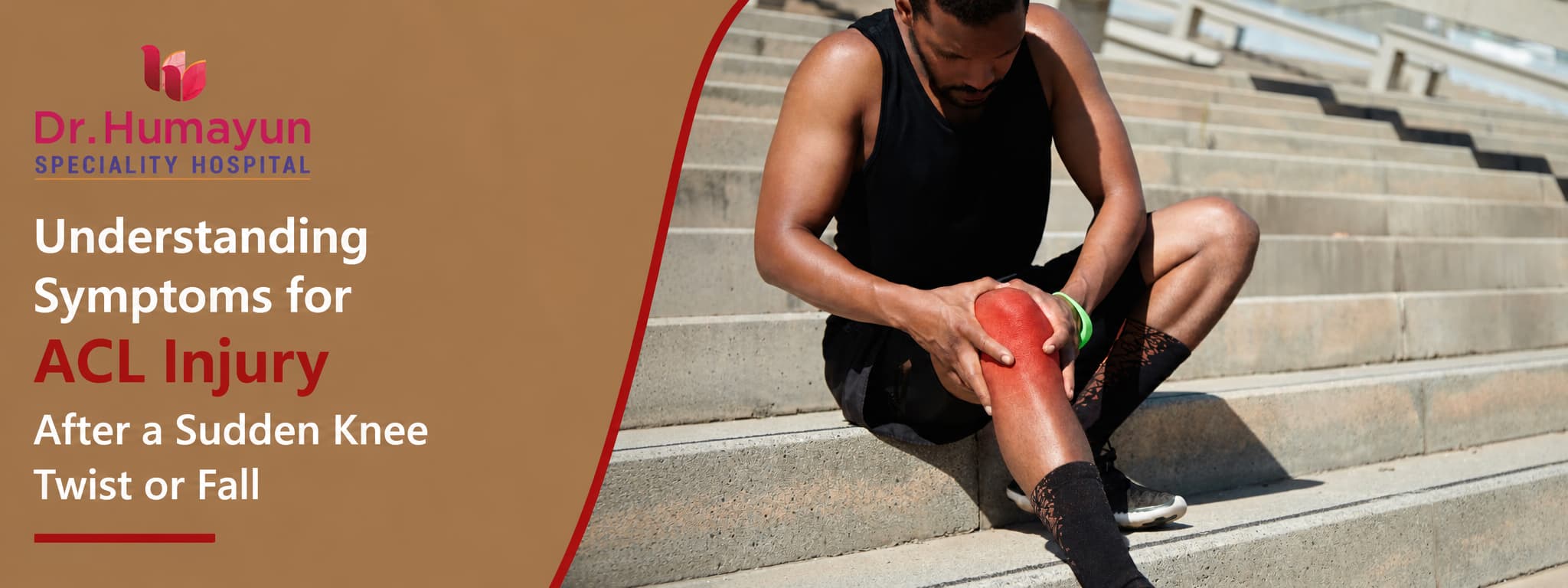

One moment you are moving normally, and the next, a sudden knee twist or an awkward landing changes everything. Experiencing a sudden trauma to the knee is alarming, but understanding exactly what occurred inside the joint is the first critical step toward a successful clinical recovery.
The Anterior Cruciate Ligament (ACL) acts as the primary stabilizing bridge inside your knee, preventing the shin bone from sliding forward and keeping the joint stable during sharp, rotational movements. When this ligament stretches or tears during a fall or sports injury, it compromises your entire physical mobility.
In urban centers like Chennai, where recreational sports and active lifestyles are growing rapidly, knowing how to spot an acute ligament injury can prevent long-term joint degradation. This guide breaks down the defining symptoms of an ACL tear, details the precise physical tests used during an orthopedic examination, and outlines the modern treatment and recovery pathways available to help you confidently regain your mobility.
What the ACL Does and Why It Matters
The anterior cruciate ligament (ACL) is one of four major ligaments inside the knee joint. Running diagonally through the centre of the knee, it connects the femur (thigh bone) to the tibia (shin bone) and performs two critical functions: it prevents the tibia from sliding forward on the femur, and it controls rotational stability of the knee during pivoting, cutting, and directional changes.
Without an intact ACL, the knee cannot generate the same resistance to rotational forces that athletic and everyday movements demand. As UF Health explains, most people with a torn ACL find their knee is unstable and may "give out" with physical activity, and those who remain untreated are at significantly higher risk of further structural damage inside the joint.
ACL injuries constitute almost half of all knee injuries and are among the most impactful orthopaedic injuries affecting active individuals. According to research published in NCBI, the mean return-to-play time following ACL reconstruction is 12.2 months, and 20% of those who sustain a complete rupture do not return to their previous sport at all. These figures reflect just how disruptive an untreated or poorly managed ACL injury can be.
Understanding the symptoms for ACL injury from the exact moment of the twist or fall is the most important step toward timely diagnosis and the best possible recovery.
The Three Grades of ACL Injury
Not every knee twist causes a complete tear. ACL injuries are classified into three grades based on the degree of ligament damage.
-
Grade 1 (Mild Sprain): The ACL fibres are stretched but remain intact and structurally continuous. The ligament is painful and tender, but knee stability is maintained. Swelling is mild. Most Grade 1 injuries recover fully with conservative management within 2 to 4 weeks.
-
Grade 2 (Partial Tear): The ACL is partially torn, with some fibres disrupted. This grade is less common than Grade 1 or Grade 3. There is more swelling and pain than in a - Grade 1, and some degree of knee instability may be present. Management depends on the individual's activity level, age, and the extent of partial disruption.
-
Grade 3 (Complete Rupture): The ACL is completely torn, either at its mid-substance or at its attachment to the bone. The ligament no longer provides rotational or anterior stability to the knee. This is the grade most commonly associated with the classic "pop" sensation, immediate swelling, and the feeling that the knee has given way. As New York Presbyterian confirms, about half of all ACL injuries include damage to other knee structures such as the articular cartilage, meniscus, or other ligaments, making early and thorough assessment critical.
Symptoms for ACL Injury: What You Feel at the Exact Moment of Injury
The moment of ACL injury is usually unmistakable. The specific combination of sensations is so characteristic that clinicians rely on this history as a primary diagnostic indicator, even before imaging.
-
The "pop" or "snap": This is the hallmark symptom for ACL injury reported by the majority of patients with a complete tear. It is described as an audible or felt crack, snap, or pop from inside the knee at the exact moment of the twist or landing. The sound is produced by the sudden rupture of the ligament under excessive tensile load. As the AAOS clinical guidelines confirm, a history of hearing or feeling a popping sensation is important in predicting an ACL injury and is one of the most consistent features in clinical history-taking.
-
Immediate pain: A sharp, sudden pain inside the knee follows the pop, often severe enough to cause the person to fall or immediately stop weight-bearing. Pain may be felt across the entire joint rather than in one specific spot.
-
Sudden loss of knee control: Many patients describe the immediate sensation that the knee "buckled," "went out," or felt completely unreliable. This reflects the sudden loss of ligamentous stability at the moment of rupture.
-
Inability to continue activity: Unlike minor knee strains, a complete ACL tear almost always ends the activity immediately. Athletes rarely "play through" a Grade 3 ACL injury.
Symptoms in the Hours and Days After the Injury
The hours following an ACL tear reveal a specific progression of symptoms that distinguish it from simpler knee injuries.
-
Rapid swelling (haemarthrosis): Significant swelling developing within 1 to 2 hours of the injury is one of the most clinically reliable indicators of a serious intra-articular injury such as an ACL tear. This rapid swelling is caused by bleeding into the joint from torn ligament blood vessels. As Cleveland Clinic confirms, an ACL tear causes pain and can keep you from being active. The knee becomes visibly larger and feels tight and warm.
-
Reduced range of motion: The combination of pain, swelling, and the protective muscle spasm that follows a significant joint injury limits how far the knee can bend or straighten in the hours after injury.
-
Ongoing instability and "giving way": As the acute pain begins to settle over the following days, the functional instability of the knee becomes more apparent. The knee may give way or feel unreliable during straightforward activities such as walking on uneven ground, descending stairs, or attempting to change direction. This instability is the defining functional consequence of ACL insufficiency.
-
Tenderness along the joint line: Tenderness is often felt along the inner (medial) or outer (lateral) joint line of the knee, particularly when an associated meniscal or cartilage injury is present alongside the ACL tear.


Why Women Face a Significantly Higher ACL Injury Risk
This section addresses a clinically important and often underrecognised fact: women are at significantly higher risk of ACL injury than men participating in the same sports and activities.
As Johns Hopkins Medicine explains, women's joints including the knee generally have more looseness and range of motion than men's. The structure of the knee joint in women plays a big role in putting them at a higher risk for an ACL tear. Research published in NCBI indicates female athletes have between 2 and 8 times the risk of suffering ACL injuries compared to male counterparts performing similar training.
The reasons are multifactorial. Women tend to have a wider pelvis relative to their leg length, creating a greater valgus angle at the knee (knees angling inward) during landing and pivoting. Women generally have greater joint laxity due to hormonal differences. Historically, women's athletic training has placed less emphasis on neuromuscular strength training, particularly hamstring and core strength, which are key to dynamic knee stabilisation. Women also tend to be more quad-dominant, placing relatively more demand on the ACL during deceleration and landing.
Understanding this elevated risk is important for prevention, for earlier investigation of knee symptoms in active women, and for realistic counselling about recovery expectations following injury.
The Mechanisms That Cause ACL Tears After a Twist or Fall
Knowing exactly how the ACL tears helps explain both the symptom pattern and the prevention strategies.
Approximately 55% of ACL injuries are non-contact, meaning they occur without an external force being applied to the knee. The most common non-contact mechanisms include:
-
Sudden deceleration and change of direction: Cutting sharply to one side while running causes enormous rotational and anterior stress on the ACL, particularly when the foot is planted and the knee is near full extension.
-
Landing from a jump: Landing with the knee straight or in a valgus (inward-collapsing) position rather than in a flexed, aligned position multiplies the forces transmitted through the ACL. Awkward landings from jumps in sports such as basketball, badminton, and volleyball are a frequent ACL injury mechanism.
-
Sudden knee twist during a fall: A fall where the body rotates while the foot remains planted on the ground generates a combination of rotational and translational forces that can rupture the ACL without any external contact.
Contact mechanisms, where an external force is applied directly to the knee from a tackle or collision, account for the remaining 45% of injuries and are more difficult to prevent through training alone.
How ACL Injury Is Diagnosed Accurately
Accurate diagnosis requires both clinical examination and targeted imaging. Neither alone is sufficient for reliable management planning.
- Clinical history: The clinician takes a detailed account of the mechanism of injury, the presence of the "pop" sensation, the speed and degree of swelling, and the functional behaviour of the knee since injury. This history alone significantly increases the probability of an ACL tear diagnosis before any tests are performed.
Physical examination including specific clinical tests:
The Lachman test is the most accurate single clinical test for ACL integrity. With the knee slightly flexed, the examiner holds the thigh stable and moves the tibia forward. Excessive forward translation of the tibia on the femur with a soft endpoint indicates ACL insufficiency. As New York Presbyterian confirms, physicians use the Lachman test as the most accurate method for determining a torn ACL.
The anterior drawer test applies anterior force to the tibia with the knee at 90 degrees and assesses for abnormal forward movement.
The pivot shift test assesses rotational instability by reproducing the subluxation and reduction of the tibiofemoral joint during rotation, confirming ACL functional insufficiency.
MRI (Magnetic Resonance Imaging): MRI is the gold standard imaging investigation for ACL injury. As New York Presbyterian confirms, MRI is the primary imaging study used to diagnose ACL injuries and can also identify additional injuries sustained alongside a torn ACL, including meniscal tears, bone bruising, cartilage damage, and associated ligament injuries.
X-ray: While X-ray does not visualise soft tissue structures like the ACL, it is performed to exclude bony avulsion fractures, particularly in younger patients where the ACL can pull a fragment of bone away from its tibial attachment.
Treatment: Non-Surgical vs Surgical
Treatment for ACL injury is not a single pathway. The right approach depends on the grade of injury, the patient's age and activity level, the presence of associated injuries, and the individual's expectations for return to sport or activity.
Non-surgical management is appropriate for Grade 1 and some Grade 2 injuries, for older or less active individuals with Grade 3 tears who do not engage in pivoting or cutting sports, and for those whose instability is well-controlled with a structured rehabilitation programme. Non-surgical management includes a structured RICE protocol immediately after injury (Rest, Ice, Compression, Elevation), followed by progressive physiotherapy focused on quadriceps and hamstring strengthening, proprioception training, and functional stability exercises. A functional knee brace may be used to support the knee during the rehabilitation period.
Surgical management (ACL reconstruction) is typically recommended for active individuals wishing to return to sport involving pivoting, cutting, or jumping; for Grade 3 tears with significant instability affecting daily life; and for injuries associated with meniscal or cartilage damage requiring simultaneous surgical repair.
As Stony Brook Medicine confirms, ACL reconstruction involves replacing the torn ligament with a graft (a piece of healthy tissue) usually taken from the patient's own body, such as the hamstring or patellar tendon, or from a donor. The new ligament acts as a scaffold while the body heals and restores stability. ACL reconstruction is an arthroscopic procedure performed through small incisions, significantly reducing recovery time compared to open surgery.
ACL Reconstruction Recovery: What to Expect
Recovery from ACL reconstruction is a structured, phased process. A realistic understanding of the timeline prevents the most common reason for re-injury: returning to activity before the reconstructed ligament has matured sufficiently.
-
Weeks 1 to 2: Focus is on controlling pain and swelling, restoring knee extension, and beginning early quadriceps activation. Most patients use crutches for the first 1 to 2 weeks.
-
Weeks 2 to 6: Progressive range of motion exercises, closed-chain strengthening, and walking without crutches. Return to driving typically occurs at this stage for right-leg injuries.
-
Months 2 to 4: Running is reintroduced on flat surfaces after confirming quadriceps strength symmetry. Proprioception and balance training become increasingly central to the programme.
-
Months 4 to 9: Sport-specific training, agility drills, and plyometric exercises are introduced progressively. Return-to-contact sport typically occurs no earlier than 9 months post-reconstruction.
Research consistently shows that 25 to 35% of athletes face re-injury within 2 to 5 years following ACL reconstruction, making the quality and duration of rehabilitation, not the surgery itself, the primary determinant of long-term outcomes.
Prevention: Reducing Your ACL Injury Risk
For athletes, recreational players, and active individuals, targeted prevention programmes significantly reduce ACL injury incidence:
-
Neuromuscular training: Exercises developing the hamstring and gluteal muscle strength that dynamically protect the ACL during landing and cutting. The FIFA 11+ and similar structured warm-up programmes have demonstrated meaningful reductions in ACL injury rates in team sports.
-
Landing mechanics: Training to land from jumps with the knees flexed and aligned over the toes, rather than straight-legged or in a valgus position, directly reduces the peak forces transmitted through the ACL.
-
Core and hip strengthening: A stronger core and stronger hip abductor muscles reduce dynamic knee valgus during single-leg loading, one of the primary mechanisms of non-contact ACL injury.
-
Progressive training loads: Avoiding sudden increases in training intensity, particularly after periods of rest, reduces the fatigue-related neuromuscular deterioration that increases ACL injury risk during the later stages of a match or training session.
Orthopaedic and Sports Medicine Care at Dr. Humayun Speciality Hospital, T. Nagar, Chennai
At Dr. Humayun Speciality Hospital, the orthopaedic team provides comprehensive assessment and management of ACL injuries for athletes, recreational players, and anyone who has experienced a significant knee twist or fall.
Services include clinical knee examination using the Lachman, anterior drawer, and pivot-shift tests; in-house MRI for complete characterisation of the ACL and associated knee structures; structured conservative rehabilitation programmes; and arthroscopic ACL reconstruction surgery for candidates requiring surgical stabilisation. Post-operative physiotherapy and functional return-to-sport protocols are an integral part of the service, not an afterthought.
The team's approach ensures that every ACL injury is accurately graded, every associated injury is identified, and every treatment decision is made with full consideration of the individual's activity goals and recovery expectations.
A Twist Does Not Have to Define Your Knee's Future
An ACL injury is serious but manageable. With an accurate diagnosis, the right treatment for your grade of injury and your lifestyle, and a properly structured rehabilitation programme, the majority of people with ACL injuries return to full activity. The key is not to dismiss the moment your knee changed, and not to wait.
Injured your knee in a sudden twist or fall and unsure whether the ACL is involved?
Chat with our care assistant for quick guidance and support and book an orthopaedic assessment at Dr. Humayun Speciality Hospital, T. Nagar, Chennai.